A Promising Breakthrough for Better Understanding and treatment of Muscle Diseases
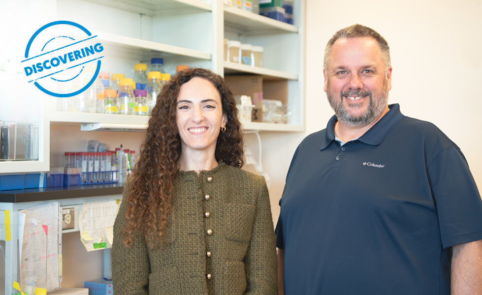
Doctoral student Sarah Nahlé and Dr Jean-François Côté
Scientists at the Montreal Clinical Research Institute (IRCM), in collaboration with partners from France and Switzerland, made a major breakthrough in understanding skeletal muscle formation. This work could pave the way for new therapeutic approaches to degenerative muscle diseases that affect thousands of people and jeopardize their autonomy.
This project was led by Sarah Nahlé, PhD student in Dr. Jean-François Côté’s laboratory, in collaboration with researchers from the IRCM, the University of Geneva, the University of Strasbourg, and Claude Bernard Lyon 1 University.
In Depth
The skeletal muscles that enable movement are formed very early in life from stem cells that become specialized cells called myocytes. These myocytes then fuse to create long muscle fibers capable of contraction. However, until now, the early stages of this process remained poorly understood.
Using cutting-edge technology called single-cell RNA sequencing, researchers were able to observe the characteristics of each cell involved in muscle formation with unprecedented precision. They discovered two distinct types of myocytes, named Mc1 and Mc2, which play complementary roles in cell fusion. These two types are distinguished by the presence of a key protein, called Myomixer, which is essential for cell fusion. Mc2 cells are present temporarily, while Mc1 cells persist throughout muscle development.
The research team also identified two important regulators, called Mef2a and Rxrg, which influence the production of Myomixer, and therefore, the ability of cells to fuse.
Why is this important?
These discoveries represent a major advance in understanding muscle development. By identifying the types of cells involved and the mechanisms that regulate them, the research team is laying the groundwork for the production of muscle cells from patients' stem cells, a crucial step towards personalized therapies. Transforming stem cells into muscle cells remains a challenge, but this work paves the way for one day creating cells capable of repairing muscles and improving the lives of people affected by muscle diseases that often remain incurable.
What's next?
The next step in this work will be to better understand how myocytes fuse to form muscle fibers. More precisely, scientists want to determine whether Mc1 and Mc2 types have distinct roles in this process and what signals direct a cell toward one type or the other. These answers will be c for laboratory reproduction of muscle formation from patient cells for therapeutic purposes.
Read full study
Acknowledgements
The work benefited from valuable support from IRCM collaborators such as Dr. Awais Javed ( Ph.D. graduate of Dr. Michel Cayouette's laboratory), Loïck Joumier (Ph.D. student in Dr. Mohan Malleshaiah's laboratory), Dr. Konstantin Khetchoumian (Research Associate in Dr. Jacques Drouin's laboratory) and Dr. Yacine Kherdjemil, head of the genome editing and disease modelling platform, as well as the teams from the IRCM's technology platforms in microscopy, bioinformatics, and histology.
The study also benefitted from partners from the University of Geneva, the University of Strasbourg, and Claude Bernard Lyon 1 University.
The project was funded by the Canadian Institutes of Health Research (CIHR), the Canada Research Chair in Cell Signaling and Cancer Metastasis, the Alain Fontaine Chair in Cancer Research of the IRCM Foundation, and the Fonds de recherche du Québec – Santé (FRQS).