An IRCM team identifies an important new mechanism
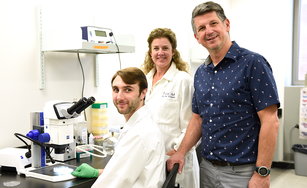
From left to right: Thomas Brown, Christine Jolicoeur, and Michel Cayouette
The Cellular Neurobiology Research Unit led by Dr. Michel Cayouette at the Montreal Clinical Research Institute (IRCM), and also Full Research Professor, Department of Medicine at Université de Montréal, has identified a key mechanism involved in the growth of nerve cells that are critical to mediate binocular vision, which allows us to see the world in 3D.
The marvel of human 3D vision
To see the world in 3D, our eyes look at an object from two different parts of the retina, a thin layer tissue at the back of the eyes that transforms light into electrical signals used by nerve cells to communicate. The overlap between these two fields of vision allows us to determine the depth, distance and speed of an object and make fast, sometimes life saving decisions. Crucial for this process is the proper growth of nerves from the eye to the brain. When these nerve cells called retinal ganglion cells send projections to the brain via the optic nerve, they either remain on the same side or cross over to the other half of the brain. It is the balance of these projections that allows us to see the world in 3D, but how exactly this is controlled remains poorly understood.
The work from the IRCM group published in the journal Cell Reports provides an important advance towards solving this mystery.
In depth
In the study, the team of scientists identified a gene called Pou3f1 that acts as a major regulator controlling the expression of dozens of other genes, which together generate the full instructions to ensure retinal ganglion cells send projections that cross to the opposite hemisphere of the brain. Furthermore, the team showed that expression of Pou3f1 in retinal stem cells is sufficient to force them to become retinal ganglion cells sending projections to the optic nerve.
“Our work identifies Pou3f1 as a critical regulator of processes underlying binocular vision in mammals and as a potential candidate for the regeneration and repair of the visual system” said Thomas Brown, a current doctoral student in the Cayouette lab and co-first author of the study with Michel Fries, a previous student in the team who recently graduated.
“Nerves are roads for information, and if they cannot send information to the appropriate area in the brain, then it leads to serious problems, as observed in blinding eye diseases like glaucoma” added Christine Jolicoeur, a senior research assistant in the team and co-author of the study.
“Our work helps understand how the roadmap of visual information is constructed and sheds light on how nerves reach the right area of the brain, which is essential information to help develop regenerative approaches for various neurodegenerative diseases”, concluded Dr. Cayouette.
Acknowledgement
This work published in Cell Reports was funded by the Canadian Institutes of Health Research.