Therapies to target neuromuscular disorders affecting million of people worldwide are on the horizon thanks to research at the Montreal Clinical Research Institute of Montreal (IRCM).
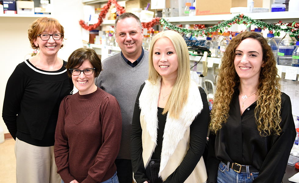
From left to right : Marie Kmita, Amélie Robert, Jean-François Côté, Viviane Tran and Sarah Nahlé.
Neuromuscular disorders affect millions of people worldwide. Now a discovery made at the Montreal Clinical Research Institute of Montreal (IRCM) opens the door to the development of targeted therapies.
Published in the journal Nature Communications, the development caps several years of research by doctoral student Viviane Tran under the direction of Université de Montréal medical professor Dr. Jean-François Côté, the IRCM’s president and scientific director, with international partners.
The formation of muscles, a complex process, requires the action of specialized cells, the myoblasts. In order for skeletal muscle to develop and regenerate, myoblasts must align with each other, move towards each other, and touch each other until their membranes are joined. This is called the myoblast fusion stage and is the basis for the formation of muscle fibers.
During embryogenesis, myoblast fusion is crucial, with mutations in certain genes resulting in the extremely rare clinical myopathy called Carey-Fineman-Ziter syndrome.In adults, an army of satellite cells is responsible for muscle growth and regeneration. In response to activation signals, satellite cells proliferate, differentiate and fuse to repair damaged myofibers. The proteins and signaling pathways that control this fusion are still being identified.
‘We didn’t think it possible’
“Until recently, myoblast fusion was the subject of only basic research," said Dr. Côté.
"We weren't interested in it in the context of disease; we didn't think it was possible to use this process to cure certain diseases. Yet, understanding in detail all the factors involved in this fusion could contribute to the development of targeted therapies."
In what was a key experiment, the researchers created a mouse model in which a protein involved in fusion is expressed in its active form in the mammal. During muscle development and regeneration, an increase in myoblast fusion was observed.
"We also observed that this mouse model, when crossed with a mouse modeling limb-girdle muscular dystrophy 2B, can improve disease phenotypes," said Tran.
Direct evidence of usefulness
The new data therefore provide direct evidence that the myoblast fusion process could be exploited for regenerative purposes and to improve the outcome of muscle diseases.
In the long term, this research shows, increasing cell fusion could "repair" muscles in other types of muscular dystrophy, such as Duchenne (occurring in 1 in 4,000 boys) or other severe conditions, such as cachexia (secondary muscle breakdown due to various diseases and some forms of cancer).
The potential to manipulate the myoblast fusion step will undoubtedly be the subject of future studies, said the researchers, who worked with colleagues at UdeM’s Institute for Research in Immunology and Cancer) and the IRCM, in Montreal and internationally.
Funding was provided by the Canadian Institutes of Health Research, the Canada Research Chairs Program, the Fonds de recherche du Québec - Santé, and the IRCM Foundation.
About this study
“Biasing the conformation of ELMO2 reveals that myoblast fusion can be exploited to improve muscle regeneration,” by Viviane Tran et al, was published Nov. 18, 2022 in Nature Communications. Funding was provided by the Canadian Institutes of Health Research, the Canada Research Chairs Program, the Fonds de recherche du Québec - Santé, and the IRCM Foundation. Collaborators from the Institut de recherche en immunologie et en cancérologie from Université de Montréal and the IRCM in Montreal and abroad also took part in this research.